1-It appears as a hypo intense lesion in T1 and hyper intense lesion in T2 in deep and sub cortical white matter with edges resemble that of vasogenic edema ( finger like ), with no definite mass effect or shift of the mid line structures.
2-Examples as HIV and Herpes simplex encephalitis.
3- Other D.D.:
-Adrenoleucodystrophy (A).
-Tuberculoma (T).
-Radiation injury (R). R E D A T
-Deep white matter ischemia (D).
-Encephalomalacia (E).
4-Adrenoleucodystrophy:
Bilateral, symmetrical coming out from ventricles.

5-Encephalitis and tuberculoma:


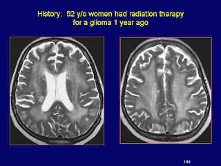
6-Radiation injury:
7-Encephalomalacia:
As the mass effect resolves and the infarcted tissue is resorbed, the adjacent sulci and ventricle will enlarge. The end result is a chronic infarct with focal areas of cystic encephalomalacia and some surrounding parenchymal change due to gliosis.

2-Examples as HIV and Herpes simplex encephalitis.
3- Other D.D.:
-Adrenoleucodystrophy (A).
-Tuberculoma (T).
-Radiation injury (R). R E D A T
-Deep white matter ischemia (D).
-Encephalomalacia (E).
4-Adrenoleucodystrophy:
Bilateral, symmetrical coming out from ventricles.

5-Encephalitis and tuberculoma:


6-Radiation injury:

7-Encephalomalacia:
As the mass effect resolves and the infarcted tissue is resorbed, the adjacent sulci and ventricle will enlarge. The end result is a chronic infarct with focal areas of cystic encephalomalacia and some surrounding parenchymal change due to gliosis.

No comments:
Post a Comment